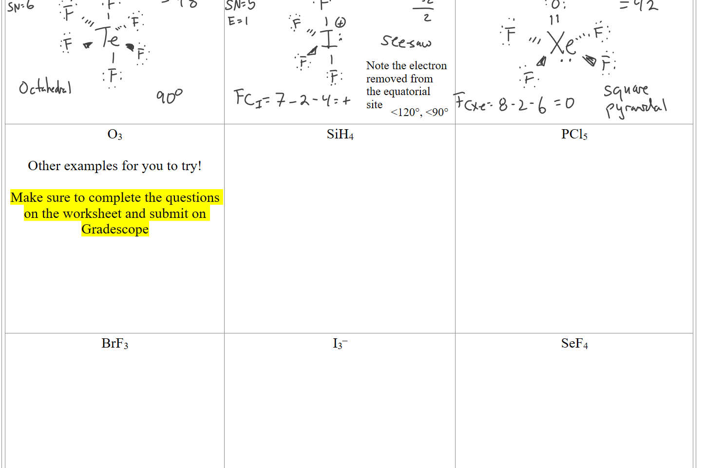
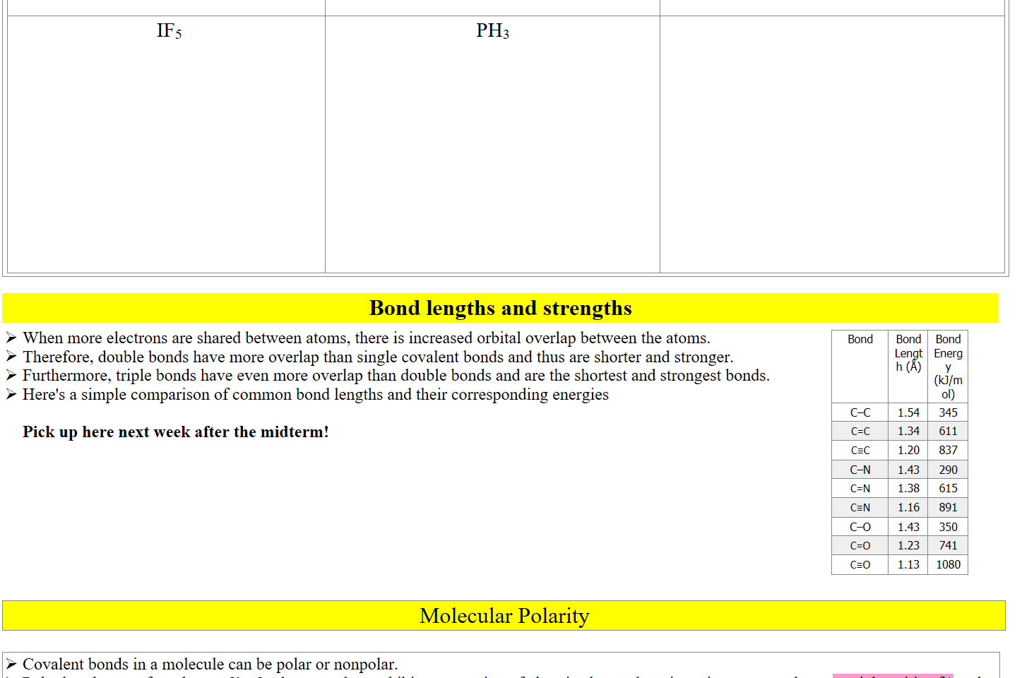
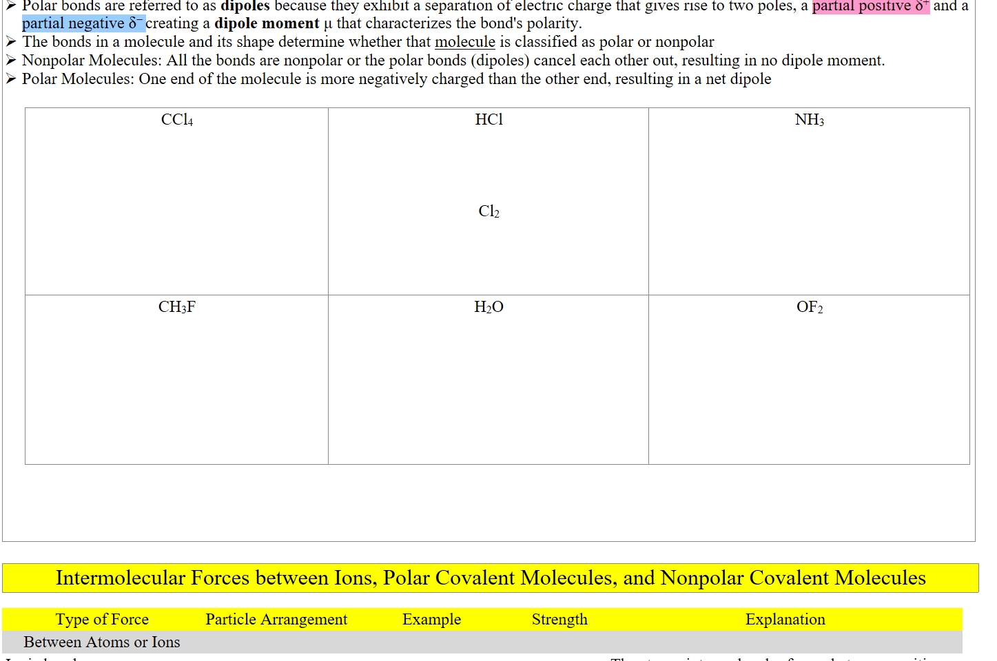
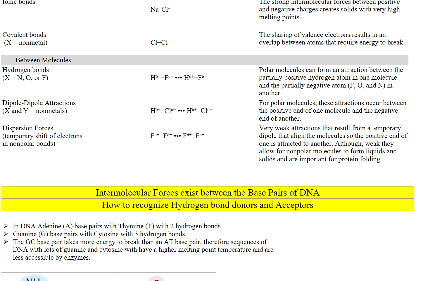
Lecture 13 - VSEPR, Molecular Polarity, Intermolecular Forces

"The basic postulate of the VSEPR model is that the arrangement of the electron pairs in a valence shell is that which places them as far apart as possible" - Ronald Gillespie Chem. Soc. Rev., 1992, 21, 59-69
Class notes for Lecture 1-12: https://bricejurban.github.io/CHEM111/
Assignments this week:
﷟HYPERLINK "https://boisestatecanvas.instructure.com/courses/28699/assignments/998317"HW 6 - Periodic Trends and Ionic Bonding (Fri 2/23)
﷟HYPERLINK "https://boisestatecanvas.instructure.com/courses/28699/assignments/998318"HW 7 - Lewis Structures (Mon 2/26)
Reading for this week: Chapter 7.5-7.10, Chapter 8
﷟HYPERLINK "https://boisestatecanvas.instructure.com/courses/28699/modules/items/3002842"Midterm 2 Practice Exam
Assignments next week:
﷟HYPERLINK "https://boisestatecanvas.instructure.com/courses/28699/assignments/945547"Midterm 2 (Tuesday (2/27) in-class)
﷟HYPERLINK "https://boisestatecanvas.instructure.com/courses/28699/assignments/993257"Emerging Tech in Chemistry (Friday (3/1) w/ replies by Sunday (3/3)

Office Hours: 
Friday 11-1 CIC 
﷟HYPERLINK "https://calendly.com/bricejurban/office-hours"By appointment

Midterm 2 Topics
Nomenclature
Ionic Compounds
Periodic Trends
Radius
Ionization Energy
Quantum Calculations
Quantum numbers
Electron Configurations
Ions, transition metals
Lewis structures
Formal Charges
Resonance
Octet Exceptions
Today's Schedule:
Tuesday (2/22)
VSEPR
Molecular Polarity
Intermolecular Forces
Looking Ahead
Tuesday (2/27) Schedule
Midterm 2 on chapters 4-7
Paper and Pencil Exam (no multiple choice)
Bring:
﷟HYPERLINK "https://boisestatecanvas.instructure.com/courses/28698/files/13892147?wrap=1"Periodic Table
﷟HYPERLINK "https://boisestatecanvas.instructure.com/courses/28698/files/14221849?wrap=1"Periodic Chart of the ions,
﷟HYPERLINK "https://boisestatecanvas.instructure.com/courses/28699/modules/items/2977345"Quantum Equations
﷟HYPERLINK "https://boisestatecanvas.instructure.com/courses/28699/modules/items/3004107"Atomic Orbital Handout
One single-sided 8.5" x 11" sheet of notes
Calculator


 Molecules exist in three-dimensions dictated by electronic repulsion

Valence-Shell Electron Pair Repulsion (VSEPR) (Gillespie-Nyholm) Theory


220px-Ronald_Gillespie.gif 
Sir-Ronald-Sydney-Nyholm.jpg Sir Ronald Sidney Nyholm (1917–1971) | Art UK
220px-Ronald_Gillespie.gif 
Sir-Ronald-Sydney-Nyholm.jpg Sir Ronald Sidney Nyholm (1917–1971) | Art UK
VSEPR theory is used to predict the shape of molecules based on the idea that electron pairs around a central atom will arrange themselves as far apart as possible to minimize repulsion.
Consider both bonding (shared between atoms) and lone (non-bonding) pairs of electrons in the valence shell of the central atom.
The geometric arrangement of the electron pairs determines the shape of the molecule.
Lone pair-lone pair > Lone pair-bonding pair > Bonding pair-bonding pair. This means lone pairs repel more strongly than bonding pairs.
Presence of lone pairs can alter the ideal bond angle and shape due to their greater repulsion. For example, NH₃ is trigonal pyramidal, not planar.
To predict a molecule's shape, draw its Lewis structure, determine the number of bonded atoms and number of nonbonding electron pairs. Use this table to help you arrange them to minimize repulsion in order to deduce the molecular geometry.
https://cms.gutow.uwosh.edu/gutow/VSEPR_TUTORIAL/shtml%20version/VSEPR_model.shtml (Visualizations of each shape)
﷟HYPERLINK "https://en.wikipedia.org/wiki/VSEPR_theory#AXE_method"VSEPR theory - Wikipedia (Helpful information about the theory)
Electron Groups
(Steric Number SN)
(Bonded Atoms +  
Number of Lone Pairs)
Electron
Geometry
Bonded Atoms
(Coordination Number)
Number of 
Nonbonding Lone Pairs
Molecular Geometry (Molecular Shape)

Bond 
Angle(s)*
Example Molecules
3D Shape
(Lone Pairs in Orange or Yellow)
2
Linear
2
0
Linear
180°
CO₂, BeCl₂
Untitled picture.png 
3
Trigonal Planar
3
0
Trigonal Planar
120°
BF₃, AlCl₃
Untitled picture.png 
3
Trigonal Planar
2
1
Bent
<120°
SO2, O3
Untitled picture.png 
4
Tetrahedral
4
0
Tetrahedral
109.5°
CH₄, SiCl₄
Untitled picture.png 
4
Tetrahedral
3
1
Trigonal Pyramidal
<109.5°
NH₃, PCl₃
Untitled picture.png 
4
Tetrahedral
2
2
Bent 
<109.5°
H₂O, H₂S
Untitled picture.png 
lone pair electrons repel more strongly
causing a distortion in bond angle
AX2
AX3
AX2E
AX4
AX3E
AX2E2
4
Tetrahedral
2
2
Bent 
<109.5°
H₂O, H₂S
Untitled picture.png 
5
Trigonal Bipyramidal
5
0
Trigonal Bipyramidal
120°, 90°
PCl₅, PF₅
Untitled picture.png 
5
Trigonal Bipyramidal
4
1
See-Saw
<120°, <90°
SF₄, SeCl₄
Untitled picture.png 
5
Trigonal Bipyramidal
3
2
T-shaped
<90°
ClF₃, BrF₃
Untitled picture.png 
5
Trigonal Bipyramidal
2
3
Linear
180°
XeF₂, I₃⁻
AX2E3-3D-balls.png undefined
6
Octahedral
6
0
Octahedral
90°
SF₆, SeF₆
Untitled picture.png 
6
Octahedral
5
1
Square Pyramidal
<90°
BrF₅, IF₅
Untitled picture.png 
6
Octahedral
4
2
Square Planar
90°
XeF₄, PtCl₄²⁻
1024px-AX4E2-3D-balls.png undefined
Electrons are removed from the equatorial sites first!
This is to reduce the number of electron repulsions
Two axial atoms and three equatorial atoms results in
two different bond angles
Water is specifically 104.5° but other atoms will be different 
AX2E2
AX5
AX4E
AX3E2
AX2E3
AX6
AX5E
AX4E2
1024px-AX4E2-3D-balls.png undefined
7 
(Rare)
Pentagonal Bipyramidal
7
0
Pentagonal Bipyramidal
90°, 72°, 180°
IF7, ReF7
800px-AX9E0-3D-balls.png undefined
8
(Rare)
Square Antiprismatic
8
0
Square Antiprismatic
109.5°, 99.6°, 70.5°
TaF83–
800px-AX9E0-3D-balls.png undefined

9
(Rare)
Tricapped Trigonal Prismatic
9
0
Tricapped Trigonal Prismatic
60°
ReH9–
800px-AX9E0-3D-balls.png undefined
*The bond angles in actual molecules may vary slightly

Rules for Finding the Molecular Geometry

Determine the correct Lewis structure for the molecule.  If it is a diatomic (has only two atoms) it is linear.  If it has 3 or more atoms continue with step 2.
Count the number of electron groups around the central atom.  A group of electrons is a bond or a nonbonding electron pair.  Each triple or double bond counts as only one group for the purposes of this model.
Based on this number of groups around the central atom the molecule falls into one of six basic categories.  Within each category there are a number of different names for the shapes depending upon the number of atoms and nonbonding groups around the central atom.
Class number (AXE notation): A = central atom X = atom attached to central atom E = nonbonding electron group on central atom


 TeF6


IF4+

XeOF4
Ink Drawings
Ink Drawings
Ink Drawings
Ink Drawings
Ink Drawings
Ink Drawings
Ink Drawings
Ink Drawings
Ink Drawings
Ink Drawings
Ink Drawings
Ink Drawings
Ink Drawings
Ink Drawings
Ink Drawings
Ink Drawings
Ink Drawings
Ink Drawings
Ink Drawings
Ink Drawings
Ink Drawings
Ink Drawings
Ink Drawings
Ink Drawings
Ink Drawings
Ink Drawings
Ink Drawings
Ink Drawings
Ink Drawings
Ink Drawings
Ink Drawings
Ink Drawings
Ink Drawings
Ink Drawings
Ink Drawings
Ink Drawings
Ink Drawings
Ink Drawings
Ink Drawings
Ink Drawings
Ink Drawings
Ink Drawings
Ink Drawings
Ink Drawings
Ink Drawings
Ink Drawings
Ink Drawings
Ink Drawings
Ink Drawings
Ink Drawings
Ink Drawings
Ink Drawings
Ink Drawings
Ink Drawings
Ink Drawings
Ink Drawings
Ink Drawings
Ink Drawings
Ink Drawings
Ink Drawings
Ink Drawings
Ink Drawings
Ink Drawings
Ink Drawings
Ink Drawings
Ink Drawings
Ink Drawings
Ink Drawings
Ink Drawings
Ink Drawings
Ink Drawings
Ink Drawings
Ink Drawings
Ink Drawings
Ink Drawings
Ink Drawings
Ink Drawings
Ink Drawings
Ink Drawings
Ink Drawings
Ink Drawings
Ink Drawings
Ink Drawings
Ink Drawings
Ink Drawings
Ink Drawings
Ink Drawings
Ink Drawings
Ink Drawings
Ink Drawings
Ink Drawings
Ink Drawings
Ink Drawings
Ink Drawings
Ink Drawings
Ink Drawings
Ink Drawings
Ink Drawings
Ink Drawings
Ink Drawings
Ink Drawings
Ink Drawings
AX7
AX8
AX9


O3

Other examples for you to try!

Make sure to complete the questions on the worksheet and submit on Gradescope


SiH4
PCl5
BrF3 


I3–
SeF4
Ink Drawings
Ink Drawings
Ink Drawings
Ink Drawings
Ink Drawings
Ink Drawings
Ink Drawings
Ink Drawings
Ink Drawings
Ink Drawings
Ink Drawings
Ink Drawings
Ink Drawings
Ink Drawings
Ink Drawings
Ink Drawings
Ink Drawings
Ink Drawings
Ink Drawings
Ink Drawings
Ink Drawings
Ink Drawings
Ink Drawings
Ink Drawings
Ink Drawings
Ink Drawings
Ink Drawings
Ink Drawings
Ink Drawings
Ink Drawings
Ink Drawings
Ink Drawings
Ink Drawings
Ink Drawings
Ink Drawings
Ink Drawings
Ink Drawings
Ink Drawings
Ink Drawings
Ink Drawings
Ink Drawings
Ink Drawings
Ink Drawings
Ink Drawings
Ink Drawings
Ink Drawings
Ink Drawings
Ink Drawings
Ink Drawings
Ink Drawings
Ink Drawings
Ink Drawings
Ink Drawings
Ink Drawings
Ink Drawings
Ink Drawings
Ink Drawings
Ink Drawings
Ink Drawings
Ink Drawings
Ink Drawings
Ink Drawings
Ink Drawings
Ink Drawings
Ink Drawings
Ink Drawings
Ink Drawings
Ink Drawings
Ink Drawings
Ink Drawings
Ink Drawings
Ink Drawings
Ink Drawings
Ink Drawings
Ink Drawings
Ink Drawings
Ink Drawings
Ink Drawings
Ink Drawings
Ink Drawings
Ink Drawings
Ink Drawings
Ink Drawings
Ink Drawings
Ink Drawings
Ink Drawings
Ink Drawings
Ink Drawings
Ink Drawings
Ink Drawings
Ink Drawings
Ink Drawings
Ink Drawings
Ink Drawings
Ink Drawings
Ink Drawings
Ink Drawings
Ink Drawings
Ink Drawings
Ink Drawings
Ink Drawings
Ink Drawings
Ink Drawings
Ink Drawings
Ink Drawings
Ink Drawings
Ink Drawings
Ink Drawings
Ink Drawings
Ink Drawings
Ink Drawings
Ink Drawings
Ink Drawings
Ink Drawings
Ink Drawings
Ink Drawings
Ink Drawings
Ink Drawings
Ink Drawings
Ink Drawings
Ink Drawings
Ink Drawings
Ink Drawings
Ink Drawings
Ink Drawings
Ink Drawings
Ink Drawings
Ink Drawings
Ink Drawings
Ink Drawings
Ink Drawings
Ink Drawings
Ink Drawings
Ink Drawings
Ink Drawings
Ink Drawings
Ink Drawings
Ink Drawings
Ink Drawings
Ink Drawings
Ink Drawings
Ink Drawings
Ink Drawings
Ink Drawings
Ink Drawings
Ink Drawings
Ink Drawings
Ink Drawings
Ink Drawings
Ink Drawings
Ink Drawings
Ink Drawings
Ink Drawings
Ink Drawings
Ink Drawings
Ink Drawings
Ink Drawings
Ink Drawings
Ink Drawings
Ink Drawings
Ink Drawings
Ink Drawings
Ink Drawings
Ink Drawings
Ink Drawings
Ink Drawings
Ink Drawings
Ink Drawings
Ink Drawings
Ink Drawings
Ink Drawings
Ink Drawings
Ink Drawings
Ink Drawings
Ink Drawings
Ink Drawings
Ink Drawings
Ink Drawings
Ink Drawings
Ink Drawings
Ink Drawings
Ink Drawings
Ink Drawings
Ink Drawings
Ink Drawings
Ink Drawings
Ink Drawings
Ink Drawings
Ink Drawings
Ink Drawings
Ink Drawings
Ink Drawings
Ink Drawings
Ink Drawings
Ink Drawings
Ink Drawings
Ink Drawings
Ink Drawings
Ink Drawings
Ink Drawings
Ink Drawings
Ink Drawings
Ink Drawings
Ink Drawings
Ink Drawings
Ink Drawings
Ink Drawings
Ink Drawings
Ink Drawings
Ink Drawings
Ink Drawings
Ink Drawings
Ink Drawings
Ink Drawings
Ink Drawings
Ink Drawings
Ink Drawings
Ink Drawings
Ink Drawings
Ink Drawings
Ink Drawings
Ink Drawings
Ink Drawings
Ink Drawings
Ink Drawings
Ink Drawings
Ink Drawings
Ink Drawings
Ink Drawings
Ink Drawings
Ink Drawings
Ink Drawings
Ink Drawings
Ink Drawings
Ink Drawings
Ink Drawings
Ink Drawings
Ink Drawings
Ink Drawings
Ink Drawings
Ink Drawings
Ink Drawings
Ink Drawings
Ink Drawings
Ink Drawings
Ink Drawings
Ink Drawings
Ink Drawings
Ink Drawings
Ink Drawings
Ink Drawings
Ink Drawings
Ink Drawings
Ink Drawings
Ink Drawings
Ink Drawings
Ink Drawings
Ink Drawings
Ink Drawings
Ink Drawings
Ink Drawings
Ink Drawings
Ink Drawings
Ink Drawings
Ink Drawings
Ink Drawings
Ink Drawings
Ink Drawings
Ink Drawings
Ink Drawings
Ink Drawings
Ink Drawings
Ink Drawings
Ink Drawings
Ink Drawings
Note the electron
removed from 
the equatorial
site
<120°, <90°
Ink Drawings
Ink Drawings

 IF5


PH3


Bond lengths and strengths
When more electrons are shared between atoms, there is increased orbital overlap between the atoms.
Therefore, double bonds have more overlap than single covalent bonds and thus are shorter and stronger.
Furthermore, triple bonds have even more overlap than double bonds and are the shortest and strongest bonds.
Here's a simple comparison of common bond lengths and their corresponding energies

Pick up here next week after the midterm!
Bond
Bond Length (Å)
Bond Energy (kJ/mol)
C–C
1.54
345
C=C
1.34
611
C≡C
1.20
837
C–N
1.43
290
C=N
1.38
615
C≡N
1.16
891
C–O
1.43
350
C=O
1.23
741
C≡O
1.13
1080

Molecular Polarity

Covalent bonds in a molecule can be polar or nonpolar.
Polar bonds are referred to as dipoles because they exhibit a separation of electric charge that gives rise to two poles, a partial positive δ+ and a partial negative δ− creating a dipole moment μ that characterizes the bond's polarity.
The bonds in a molecule and its shape determine whether that molecule is classified as polar or nonpolar
Nonpolar Molecules: All the bonds are nonpolar or the polar bonds (dipoles) cancel each other out, resulting in no dipole moment.
Polar Molecules: One end of the molecule is more negatively charged than the other end, resulting in a net dipole

CCl4


HCl


Cl2
NH3
CH3F


H2O 
OF2


Intermolecular Forces between Ions, Polar Covalent Molecules, and Nonpolar Covalent Molecules

Type of Force
Particle Arrangement
Example
Strength
Explanation
Between Atoms or Ions


Ionic bonds


The strong intermolecular forces between positive and negative charges creates solids with very high melting points.
Ionic bonds


Na+Cl–

The strong intermolecular forces between positive and negative charges creates solids with very high melting points.
Covalent bonds
 (X = nonmetal)


Cl−Cl

The sharing of valence electrons results in an overlap between atoms that require energy to break.
Between Molecules


Hydrogen bonds
(X = N, O, or F)


Hδ+−Fδ− ••• Hδ+−Fδ−

Polar molecules can form an attraction between the partially positive hydrogen atom in one molecule and the partially negative atom (F, O, and N) in another. 
Dipole-Dipole Attractions
(X and Y = nonmetals)


Hδ+−Clδ− ••• Hδ+−Clδ−

For polar molecules, these attractions occur between the positive end of one molecule and the negative end of another. 
Dispersion Forces
(temporary shift of electrons in nonpolar bonds)


Fδ+−Fδ− ••• Fδ+−Fδ−

Very weak attractions that result from a temporary dipole that align the molecules so the positive end of one is attracted to another. Although, weak they allow for nonpolar molecules to form liquids and solids and are important for protein folding


Intermolecular Forces exist between the Base Pairs of DNA
How to recognize Hydrogen bond donors and Acceptors


In DNA Adenine (A) base pairs with Thymine (T) with 2 hydrogen bonds
Guanine (G) base pairs with Cytosine with 3 hydrogen bonds
The GC base pair takes more energy to break than an AT base pair, therefore sequences of DNA with lots of guanine and cytosine with have a higher melting point temperature and are less accessible by enzymes.


Untitled picture.png Machine generated alternative text:
NH 
-N 
C 
CH 
Adenine (Ade) 
Untitled picture.png Machine generated alternative text:
CH3 
Thymine (Thy)l
Untitled picture.png Machine generated alternative text:
NH 
-N 
C 
CH 
Adenine (Ade) 

Untitled picture.png Machine generated alternative text:
CH3 
Thymine (Thy)l 
Untitled picture.png Machine generated alternative text:
CH 
142N 
uanine (Gua) 

Untitled picture.png Machine generated alternative text:
NH 
NCH 
CH 
Cytosine (Cyt) 


There are lots of hydrogen bond donors and acceptors in the base pairs of DNA
The left side of Adenine in the above image will base pair with the left side of Thymine because they can form two hydrogen bonds
Hydrogen bond donor-acceptor pair 1 above
Hydrogen bond acceptor-donor pair 2 above
The left side of Guanine in the above image will base pair with the left side of Cytosine because they can form three hydrogen bonds
Hydrogen bond acceptor-donor pair 3 above
Hydrogen bond donor-acceptor pair 4 above
Hydrogen bond donor-acceptor pair 5 above
This base pairing is shown in the image below from the Color Atlas of Biochemistry
This base pairing is only possible when the polarity of two strands of DNA are running in opposite directions. This forms a double helix structure!
Untitled picture.png Machine generated alternative text:
— A. DNA: structure 
1. Formula 
CH3 0 
o 
CH20H 
CH2 
CH2 
Minor 
groove 
2. Double strand 
Major 
groove 
OH 
2'-deoxy- 
D-ribose
Untitled picture.png Machine generated alternative text:
— A. DNA: structure 
1. Formula 
CH3 0 
o 
CH20H 
CH2 
CH2 
Minor 
groove 
2. Double strand 
Major 
groove 
OH 
2'-deoxy- 
D-ribose


Created with OneNote.